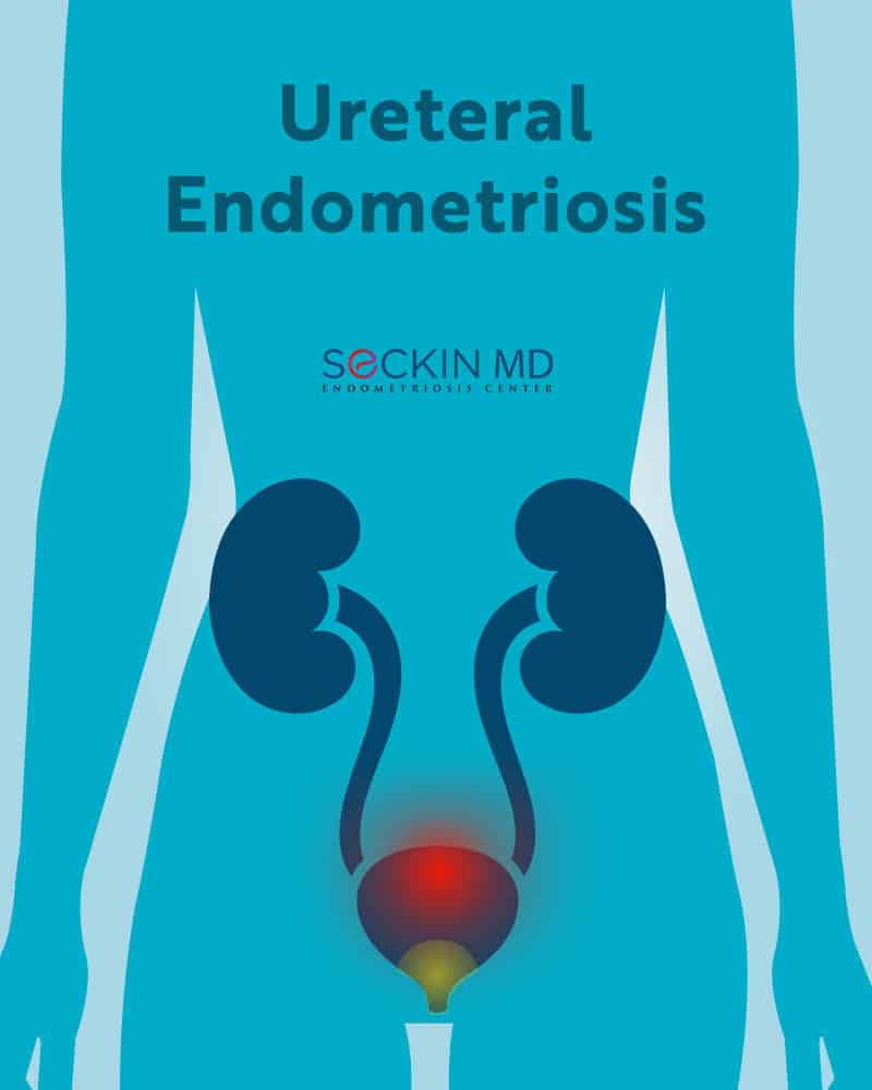
Ureteral endometriosis is a form of urinary tract endometriosis (UTE). UTE is a rare form of deep infiltrating endometriosis (DIE) in which the growth of endometriosis (tissue resembling the endometrium) occurs in the bladder, ureters, and kidneys. DIE usually develops after ovarian endometriomas spill (cyst rupture) into other parts of the abdomen and pelvis.
Ureteral endometriosis can be either extrinsic or intrinsic. Extrinsic ureteral endometriosis results from endometriosis lesions occurring outside the ureter and compressing it. Intrinsic ureteral endometriosis, which accounts for nearly 20% of cases, occurs within the wall, muscle, and inner layers of the ureter.
Prevalence
UTE affects between 0.3% and 12% of endometriosis cases and between 20% and 52.6% of those diagnosed with DIE.
UTE predominantly occurs in the bladder (85%), but it can also affect the ureter (10%), kidneys (4%), and urethra (2%).
Get a Second Opinion
Our endometriosis specialists are dedicated to providing patients with expert care. Whether you have been diagnosed or are looking to find a doctor, they are ready to help.Our office is located on 872 Fifth Avenue New York, NY 10065.
You may call us at (212) 988-1444 or have your case reviewed by clicking here.
What causes ureteral endometriosis?
The exact cause for endometriosis itself is not fully clear. This makes it all the more complex to properly explain the cause of ureteral endometriosis. The common theories used to explain the origin of ureteral endometriosis include retrograde menstruation, stem cells, and immune factors.
In some women, UTE may also be iatrogenic, resulting from previous Caesarean sections.
What are the symptoms?
UTE lesions that often occur superficially on the peritoneum do not grow into the bladder. But they can cause pain when having a full bladder or when passing urine. However, distinguishing this pain from the one that arises as a result of other forms of pelvic endometriosis is difficult.
Superficial lesions can also occur on the ureters. They can impact the functioning of nerves that control the bladder, vagina, and bowel movements.
Symptoms of UTE overlap with that of peritoneal endometriosis. Women with UTE experience pelvic pain and dysuria (pain with urination) along with frequent urinary tract infections, changes to urination frequency, hematuria (blood in the urine), and rarely, urinary incontinence. Nearly 50% of ureteral endometriosis cases, however, are asymptomatic with about 25% of women experiencing flank pain and 15% having hematuria. Other symptoms include painful periods and intercourse.
The DIE forms of UTE also cause symptoms such as pelvic pain, dysuria, and in some cases, hematuria. Ureteral endometriosis can affect the free passage of urine from the kidneys to the bladder. This can result in hydroureter (dilation of ureters due to accumulation of urine) or even hydronephrosis (enlargement of kidneys due to blockage and accumulation of urine). Both of these can lead to severe back pain and potential kidney failure without timely intervention.
Diagnosing ureteral endometriosis
Diagnosing ureteral endometriosis can be quite challenging. It is, therefore, is important to consult a surgeon who can listen to and understand your symptoms. Initial stages of diagnosis of endometriosis affecting any area include taking the patient’s medical history followed by pelvic examination and imaging techniques such as ultrasound, sonohysterography, or magnetic resonance imaging (MRI) to identify the location, and stage of the disease.
Intravenous pyelogram (IVP) is a good imaging technique to predict intrinsic forms of ureteral endometriosis. IVP also helps to evaluate ureter structure after treatment.
Transabdominal ultrasonography can help visualize ureter structure and obstruction in the pelvic region. It can also help see other potential sites of endometriosis lesions in the pelvis. When combined with transvaginal ultrasonography, it is possible to visualize the region from the anterior parametrium (the connective tissue that surrounds the uterus separating the cervix from the bladder) to the renal pelvis (upper end of the ureter).
Laparoscopic excision surgery followed by histological examination is the gold standard for confirming endometriosis in the urinary tract. The presence of other urinary tract infections and neoplasms (tumors) are excluded before confirming ureteral endometriosis.
Treatment
The aim of ureteral endometriosis treatment is to remove endometriosis lesions in the urinary tract and preserve renal function. In cases of mild ureteral endometriosis, medical management with GnRH analogs, combined oral contraceptives, progestin, and aromatase inhibitors may help. However, this is not recommended if disease progression or recurrence is expected in which case surgical methods are the best option.
Also, medical treatment alone cannot revert the fibrosis resulting from ureteral endometriosis that leads to ureter obstruction. Therefore, surgical intervention is usually necessary for both intrinsic and extrinsic forms of ureteral endometriosis.
The kidney can be preserved if it still has a greater than 10% glomerular filtration rate (GFR). GFR is a measure of how much blood passes through the kidney’s glomeruli (structures in the nephron that filter blood) per minute. A less than 10% GFR may mean that the kidney needs to be removed (nephrectomy).
Prior to any surgical procedure (as long as there is no complete ureteral obstruction), ureterolysis is performed to free the ureter from any pressure to avoid injury. The actual surgical procedure and anatomical corrections involved thereafter depend on the extent of damage, segment of the ureter involved, and renal function.
Dr. Seckin’s team is able to isolate and remove all forms of endometriosis, including the deep infiltrating forms in the ureter, using laparoscopic excision surgery for lasting relief. Using a patented Aqua Blue Contrast dye to easily identify endometriosis lesions combined with “cold excision” Dr. Seckin minimizes scarring compared to any of the other disease management methods.
Get a Second Opinion
Our endometriosis specialists are dedicated to providing patients with expert care. Whether you have been diagnosed or are looking to find a doctor, they are ready to help.Our office is located on 872 Fifth Avenue New York, NY 10065.
You may call us at (646) 960-3080 or have your case reviewed by clicking here.