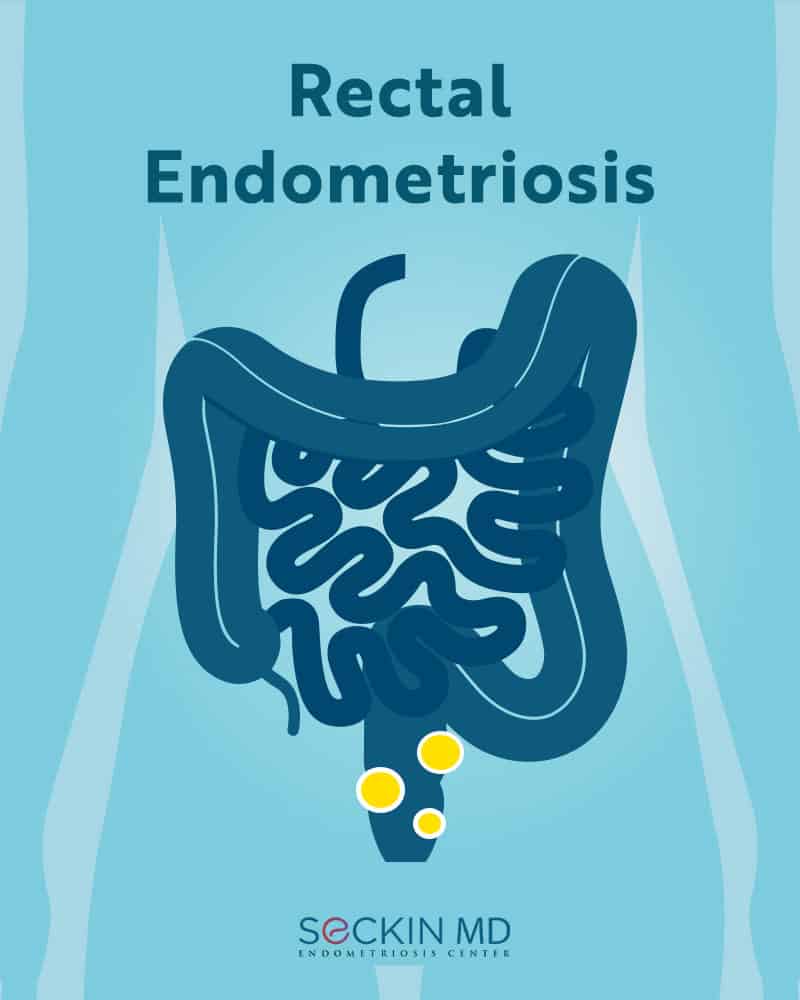
Endometriosis is a chronic, progressive disease affecting nearly 10-12% of women of reproductive age. The disease is characterized by tissue resembling the lining of the uterus growing outside the uterine cavity. This extraneous endometrial tissue may commonly occur in the peritoneum or in extra-pelvic organs such as the bowels, appendix, pancreas, and thoracic cavity. The space between the uterus and the rectum, known as the posterior cul-de-sac, is one of the common sites of endometriosis leading to rectal endometriosis.
About the rectum
The rectum is the last portion of the large intestine that converges into the anal cavity. The rectum is about 12 to 15 cm in length and serves to temporarily store feces till defecation. As such, it helps in maintaining fecal continence.
The anatomical position of the rectum is different in males and females. In women, the cervix, uterus, and vagina are present anterior to the rectum. A peritoneal layer covers the anterior and later upper third of the rectum by about six to eight cm from the point where it joins the anus.
The peritoneal layer forms the rectouterine pouch in women between the rectum and the uterus. Several diseases such as endometriosis, abscess, or hernia can affect the rectouterine pouch.
Get a Second Opinion
Our endometriosis specialists are dedicated to providing patients with expert care. Whether you have been diagnosed or are looking to find a doctor, they are ready to help.Our office is located on 872 Fifth Avenue New York, NY 10065.
You may call us at (212) 988-1444 or have your case reviewed by clicking here.
Bowel endometriosis and the rectum
Endometriosis can affect the bowels. Bowel endometriosis occurs in about 5-12% of women with endometriosis and about 90% of these cases involve the rectum. Unfortunately, doctors often ignore the endometriosis symptoms or misdiagnose them as irritable bowel disease or Crohn’s disease.
Doctors can also misdiagnose rectal endometriosis as rectal cancer. Symptoms coinciding with the onset of menstruation may be an indication of bowel endometriosis.
Normally, endometriosis only affects the outer serosal layer of the bowels. Deep-infiltrating endometriosis (DIE) lesions are usually greater than 5 mm in depth and affect the muscular layers underneath.
Causes of rectal endometriosis
It is not clear what causes bowel or rectal endometriosis. The theory of retrograde menstruation could explain the deposition of endometrial tissue outside the uterus.
Other recent theories include dysfunction in gene regulation that causes stem cells to implant and alter the immune environment around the peritoneum.
Other possible factors include oxidative stress and the presence of reactive oxygen species. These can contribute to the proliferation of endometrial tissue.
The lesions can also cause fibrotic reactions. These may lead to adhesions and anatomical distortions that can have a lasting effect on the symptoms even if the lesion itself isn’t active.
Symptoms of rectal endometriosis
The main symptoms of rectal endometriosis include digestive problems such as pain during defecation and constipation during monthly cycles. Rectal stenosis (narrowing of the rectal passage) also occurs in nearly 26.4% of women diagnosed with rectal endometriosis. Rectal stenosis may happen regardless of the presence of deep-infiltrating endometrial tissue. Symptoms often mimic those of irritable bowel syndrome or colonic adenocarcinoma.
Symptom severity may not always correlate with disease staging or lesion size. The presence of rectal endometriosis is also an indication of other forms of endometriosis such as ovarian endometrioma or retro-cervical DIE. DIE lesions can also occur together with inflammatory bowel disease, contributing to overall pain and symptom intensity.
Diagnosis of rectal endometriosis
Diagnosis of rectal endometriosis includes a physical examination of the rectum. Imaging techniques such as transvaginal ultrasound and magnetic resonance imaging (MRI) can also be useful.
Transvaginal ultrasound can offer better sensitivity (75-98%) and specificity for detecting DIE in the rectum compared to MRI, transrectal ultrasonography, or computer tomography (CT). Transvaginal ultrasound can also help in determining the size, number, and extent of invasion of these lesions, and their distance from the anus.
Apart from transvaginal ultrasound, doctors may also recommend the use of colonoscopy, double-contrast barium enema, and CT.
However, imaging techniques alone cannot offer a conclusive diagnosis. In one case study, doctors initially suspected a 36-year-old woman with rectal endometriosis of having cervical cancer. Colonoscopy did reveal a mass at the rectum about 4 cm from the anus. However, it was only after adequately sampling the recessed tissue for biopsy could the doctors confirm rectal endometriosis. Therefore, laparoscopic excision followed by biopsy and identification of appropriate histopathological markers is the only way to decisively confirm rectal endometriosis.
Sometimes, the presence of another disease can eclipse rectal endometriosis leading to misdiagnosis. In another case study of a 37-year-old woman, the preliminary diagnosis based on symptoms showed the presence of hemorrhoids. However, the patient also had a small endometrial nodule in the rectum approximately 8 cm from her anus. Doctors detected these only during the digital rectal examination.
Treatment of rectal endometriosis
Surgery is necessary to fully resect the lesions. But currently, there is no standardization on which lesions should be operated and to which extent. Surgery for rectal endometriosis can be associated with significant morbidities and complications. So, the treatment must be individualized and proper counseling of the patient is a must before committing to the procedure.
The size of the lesions and the presence of fibrotic tissue play an important role in determining the course of surgery. Doctors must perform surgery in a way to avoid significant distortion of bowel anatomy. Complete excision of fibrotic tissue is necessary to minimize the chances of disease recurrence.
Other factors that decide the course of surgery include the depth of the lesions, the extent of bowel involvement, the distance to the anus, and lymph node involvement. All these factors are not independent of each other. Thus, a multidisciplinary approach is necessary to fully assess all risks involved.
Laparoscopic deep excision surgery is the preferred method to identify and remove all rectal endometriosis lesions and the accompanying fibrous tissue. Implantation of a stoma may be considered after informing the patient if the lesions are found less than 5 cm from the anal margin.
The surgeons at Seckin Endometriosis Center (SEC) have decades of experience in laparoscopic excision surgery and are able to use innovative techniques such as the Aqua Blue Contrast (ABC)TM and cold excision to identify and excise every lesion while preserving maximum organ function. SEC surgeons led by Dr. Tamer Seckin are also able to perform reconstruction and repair of the peritoneum.
They use a multidisciplinary approach and always ensure patients are involved in key decisions. For example, we will always inform our patients if there is a need to perform bowel resection in addition to excision of rectal endometriosis tissue.
Patient story
Carolyn C. experienced what most women do when they begin seeking respite from excruciating monthly periods — a misdiagnosis. Carolyn’s case was misdiagnosed as irritable bowel syndrome and later as diverticulitis. After one endo symptom after the other started rearing its head, Carolyn realized that changing doctors from urologists to gynecologists did little to address the root cause of her pain — until she chanced upon Dr. Seckin’s resources online.
Read about how approaching Dr. Seckin changed Carolyn’s life for the better.
Get a Second Opinion
Our endometriosis specialists are dedicated to providing patients with expert care. Whether you have been diagnosed or are looking to find a doctor, they are ready to help.Our office is located on 872 Fifth Avenue New York, NY 10065.
You may call us at (646) 960-3080 or have your case reviewed by clicking here.