Dual Compartment Surgery to Treat Pneumothorax due to Endometriosis
Treat Pneumothorax due to Endometriosis
Catamenial Pneumothorax (partial or complete lung collapse during the time of menstruation) is almost always a characteristic of thoracic endometriosis. However, the condition is difficult to diagnose and multi-disciplinary expertise is necessary for its treatment.
Dr. Tamer Seckin recently presented how pores in the peritoneum due to endometriosis can lead to pneumothorax at the 9th Asian Conference on Endometriosis that took place in Colombo, Sri Lanka. Dr. Seckin advocated a dual compartment surgical approach to identify and excise endometrial lesions in the thorax and pelvic/abdominal cavities.
Endometriosis and pneumothorax
Endometriosis is essentially a disease of the pelvic peritoneum. However, it can also spread to other parts of the body.
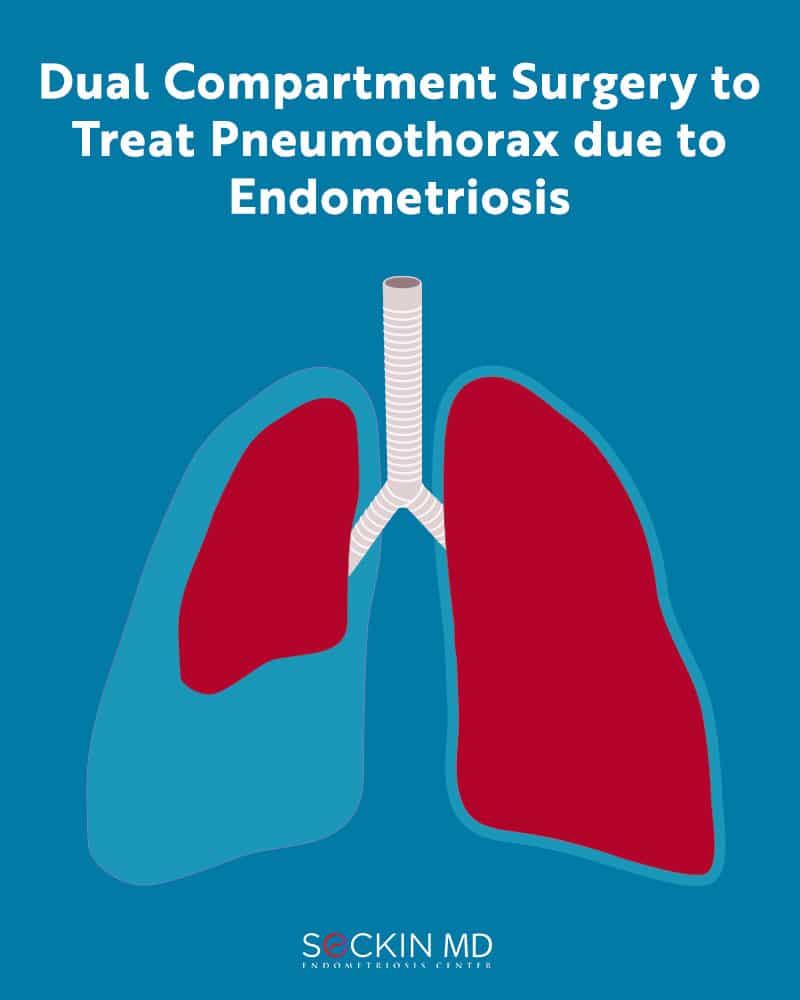
Thoracic endometriosis and diaphragmatic endometriosis are rare forms of extra-pelvic endometriosis that spread from the pelvic regions to the thorax and diaphragm, respectively. They are characterized by recurrent episodes of pneumothorax and are often not properly diagnosed.
Thoracic endometriosis can lead to symptoms such as catamenial pneumothorax (pneumothorax during menstruation), and catamenial hemothorax (blood collection in the pleural space during menstruation), catamenial hemoptysis (coughing up blood during menstruation), and lung nodules.
The first correlation between endometriosis and pneumothorax was established by Dr. Elmer Maurer and colleagues in 1958 where he described incidents of recurring pneumothorax in women coinciding with the onset of menstruation. This case was of a 35-year-old woman who had experienced 15 episodes of spontaneous pneumothorax that always coincided with menstruation.
Thoracotomy investigations revealed a 2 cm hole in the right diaphragm while subsequent laparotomy confirmed pelvic endometriosis. This led Maurer and colleagues, to conclude that the passage of air through the genital tract into the peritoneal cavity via the hole in the diaphragm and finally into the thoracic region led to lung collapse.
Physiology of pneumothorax due to endometriosis
Accumulation of air within the pleural layers of the lung can apply pressure and cause it to collapse. The normal air pressure within the pleural space of each lung is negative, about -3 to -7 mm Hg. The intra-uterine pressure during menstruation is about 20 to 56 mm Hg. Therefore, any diaphragmatic defect can cause the thorax to act as a suction pump due to the negative pressure causing air to rush from the peritoneal cavity and into the thoracic cavity.
Pneumothorax associated with endometriosis is categorized as a secondary spontaneous pneumothorax, which has a high recurrence rate and is comparatively more lethal than a primary spontaneous pneumothorax.
Aqua Blue Contrast™ technique to better understand the peritoneum
Endometriosis is primarily a peritoneal disease. So, understanding the inflammatory processes leading to surface changes in the pelvic peritoneum is the key to proper diagnosis and understanding of pelvic pain and associated symptoms such as infertility.
A normal peritoneum appears transparent or translucent and has a plain texture with a smooth and shiny surface. It shows no abnormal vascularity and has no fibrosis and no holes.
Using our patented Aqua Blue Contrast (ABC)™ technique, it is possible to identify features of the peritoneum that are not normally visible. ABC uses 5 mL of methylene blue diluted in 3 liters of Ringer’s lactate solution. In this technique, the peritoneal area under investigation is submerged in the blue dye solution and distended using hydrodistension.
This eliminates direct gas pressure on the peritoneal surfaces. It also improves visibility by removing the reflection of red and yellow lights. This way the surgeon has a much clearer view of the peritoneal layers.
ABC allows the surgeon to clearly visualize endometrial lesions and pores on the peritoneum. This minimizes the risk of damaging surrounding healthy tissue and also identifying multiple lesions for removal in one go.
Dual compartment surgery for thoracic endometriosis
Thoracoscopy and laparoscopy are two minimally invasive surgical techniques that can help the surgeon diagnose and treat thoracic endometriosis. At Seckin Endometriosis Center, we always advocate a dual compartment approach.
There is a strong correlation between endometriosis of the pelvic region and the thorax. Up to 80% of women with thoracic endometriosis have pelvic and abdominal endometrial lesions as well. Thus, it is important to properly assess the diaphragm and excise all endometrial lesions in these cavities in order to prevent the recurrence of pneumothorax and preserve fertility.
Our dual compartment approach uses both laparoscopic excision surgery, which is the gold standard for treating endometriosis, and video-assisted thoracoscopic surgery (VATS). Instead of performing them distinctly in two separate procedures, our approach involves the thoracic surgeon handling the VATS while the gynecological surgeon performs laparoscopic excision.
Proven expertise in dual compartment surgery
Between January 2013 and July 2021, surgeons at the Seckin Endometriosis Center have performed 1,664 laparoscopic endometriosis surgeries. Of these 37 patients were diagnosed with thoracic endometriosis, which constitutes roughly 2% of our surgical referrals. 18 patients had prior pneumothorax while nine had multiple pneumothoraxes. One patient even had 17 chest tubes placed.
Among these 37 patients, 67% had prior endometriosis surgery while 35% had peritoneal endometriosis. 65% of these patients also showed advanced stage endometriosis with instances of deep-infiltrating endometriosis (DIE) and ovarian endometrioma.
Lesions in the abdominal cavity can be excised and repaired via laparoscopic suturing. The lesions removed from the thoracic cavity can also be repaired via stapling. The experts at Seckin Endometriosis Center have many years of experience with these techniques including transmural suturing through the entire thickness of the diaphragm. Preemptively, we also add a mesh on the diaphragm even if there are no visible holes if we infer that there are diaphragmatic defects that could lead to pneumothorax in the future.
Get a Second Opinion
Our endometriosis specialists are dedicated to providing patients with expert care. Whether you have been diagnosed or are looking to find a doctor, they are ready to help.Our office is located on 872 Fifth Avenue New York, NY 10065.
You may call us at (646) 960-3080 or have your case reviewed by clicking here.